In a detailed clinical study, researchers at the National Institutes of Health have found differences in the brains and immune systems of people with post-infectious myalgic encephalomyelitis/chronic fatigue syndrome (PI-ME/CFS).
They also found distinct differences between men and women with the disease. The findings were published in Nature Communications.
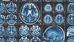
A team of multidisciplinary researchers discovered how feelings of fatigue are processed in the brains of people with ME/CFS. Results from functional magnetic resonance imaging (fMRI) brain scans showed that people with ME/CFS had lower activity in a brain region called the temporal-parietal junction (TPJ), which may cause fatigue by disrupting the way the brain decides how to exert effort.
They also analyzed spinal fluid collected from participants and found abnormally low levels of catecholamines and other molecules that help regulate the nervous system in people with ME/CFS compared to healthy controls. Reduced levels of certain catecholamines were associated with worse motor performance, effort-related behaviors, and cognitive symptoms. These findings suggest a link between specific abnormalities or imbalances in the brain and ME/CFS.
Immune testing revealed that the ME/CFS group had higher levels of naive B cells and lower levels of switched memory B cells—cells that help the immune system fight off pathogens—in blood compared to healthy controls. Naive B cells are always present in the body and activate when they encounter any given antigen, a foreign substance that triggers the immune system. Memory B cells respond to a specific antigen and help maintain adaptive or acquired immunity. More studies are needed to determine how these immune markers relate to brain dysfunction and fatigue in ME/CFS.
To study fatigue, Dr. Nath and his team asked participants to make risk-based decisions about exerting physical effort. This allowed them to assess the cognitive aspects of fatigue, or how an individual decides how much effort to exert when given a choice. People with ME/CFS had difficulties with the effort choice task and with sustaining effort. The motor cortex, a brain region in charge of telling the body to move, also remained abnormally active during fatiguing tasks. There were no signs of muscle fatigue. This suggests that fatigue in ME/CFS could be caused by a dysfunction of brain regions that drive the motor cortex, such as the TPJ.
Deeper analyses revealed differences between men and women in gene expression patterns, immune cell populations, and metabolic markers. Males had altered T cell activation, as well as markers of innate immunity, while females had abnormal B cell and white blood cell growth patterns. Men and women also had distinct markers of inflammation.
The study, which was conducted at the NIH Clinical Center, took a comprehensive look at ME/CFS that developed after a viral or bacterial infection. The team used state-of-the-art techniques to examine 17 people with PI-ME/CFS who had been sick for less than five years and 21 healthy controls. Participants were screened and medically evaluated for ME/CFS over several days and underwent extensive tests, including clinical exams, fMRI brain imaging, physical and cognitive performance tests, autonomic function tests, skin and muscle biopsies, and advanced analyses of blood and spinal fluid. Participants also spent time in metabolic chambers where, under controlled conditions, their diet, energy consumption, metabolism, sleep patterns, and gut microbiome were evaluated. During a second visit, they completed a cardiopulmonary exercise test to measure the body’s response to exercise.
The highly collaborative project involved 75 investigators across 15 institutes and centers in the NIH Intramural Research Program, and at national and international institutions.

