Breast cancer biomarkers, and a new clinical category for HER2 expression
Recent advances in treating breast cancer are reaching back to the past to the first known biomarker, human epidermal growth factor, to determine its value in treating patients with lower levels of HER2 expression and potentially open the door to new, targeted, and personalized therapies for many patients.
Estrogen receptor, progesterone receptor, and human epidermal growth factor 2 are at the cornerstone of the pathology workup for breast cancer patients and the starting point for oncologists when determining the treatment options. Biomarker status is typically determined by immunohistochemistry, with known correlations to the histologic features of the tumor. This testing may be repeated on subsequent tissue samples throughout the course of the disease, and the information is used to adjust or change the therapy in response to tumor biology and biomarker expression, which may also change over time.
Recently approved therapies are advancing the treatment and management options for breast cancer patients, building on the foundation of what we have learned about the types of breast cancer and their corresponding biomarker profiles. While pathologists have always been at the center of tissue diagnostics and patient care, the impact of the pathologist’s report and findings are just as important today. The treatment and management options available to oncologists are directly informed by the information from pathologists, so coordination and collaboration among clinical colleagues has never been more crucial.
The therapeutic advances, particularly those that are directed or informed by pathology testing, have changed the course of breast cancer for many to a chronic disease state, with patients living longer and with an improved quality of life. However, breast cancer is still the second leading cause of cancer death among women overall and the leading cause of cancer death among Hispanic women in the United States. About 42,000 women and 500 men in the United States die each year from breast cancer, with black women having a higher rate of death than white women.1
HER2-low research
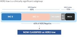
Research looking at HER2 immunohistochemistry (IHC) expression beyond the established binary categories of HER2 positive (IHC 3+ or 2+/ISH amplified) and HER2 negative (0, 1+ or 2+/ISH non-amplified) in metastatic breast cancer (mBC) patients shows that patients with HER2-low expression, defined as 1+, 2+/non-amplified also benefit from targeted HER2 therapy. The DESTINY-Breast04 clinical trial (DB-04) enrolled unresectable or mBC patients previously treated with chemotherapy.2 Both hormone receptor positive and negative patients were included, and HER2 expression was defined as 1+/never ISH or 2+/non-amplified patient who had never been reported as HER2 positive. The authors used central testing of the recent tumor tissue specimens as well as archive specimens to determine HER2 score. The primary outcome was progression-free survival and overall survival was a secondary outcome. The therapy, trastuzumab deruxtecan, nearly doubled the median progression-free survival in HR+/HER2-low patients and demonstrated anti-tumor activity. Trastuzumab deruxtecan is a HER2 directed monoclonal antibody that provides targeted delivery of the cytotoxic agent and consists of the same amino acid sequence as trastuzumab.
The findings of the DESTINY-Breast04 trial mean that the 50–60% of patients previously categorized as HER2 negative, and with 1+ and 2+/non amplified expression, comprise this new HER2-low category and are eligible for a new targeted treatment that increases survival. See Figure 1.3,4
HER2 considerations for pathologists
Pathologists should understand that DB-04 was designed using the HER2 (4B5) clone, using a locked protocol with recommended cell conditioning and antibody incubation times. The scores were made using the current ASCO/CAP guidelines.5 The reproducibility of the results in an IHC assay is critical to the accurate interpretation and reporting of results for patients.
Based on current HER2 assays and protocols in histology laboratories, there may be a need to validate for HER2/HER2-low expression. There is no proficiency testing to assess for HER2-low expression at this time. A lab resource has been established to assist in identifying HER2-low expression.6
Pathology reports should follow the recommendations for breast biomarker results and include the IHC score, the percentage of cells and the pattern of staining for those cells used to determine the score, along with the type of test and the primary antibody used.
Diagnostic tools and innovations for patients
The use of digital pathology and algorithms for biomarker interpretation and scoring is becoming more common, allowing for more accurate results and efficiencies for busy pathology practices. Molecular testing is also more common and helps to inform risk stratification based on mutations, particularly in younger patients, patients with advanced/aggressive disease, and relevant family history of breast cancer. Advances such as dual-stain in-situ hybridization (ISH) have also created efficiencies for some labs and provided an important option for reflex testing of IHC 2+ cases.
Diagnostic results, including biomarker/immunohistochemistry interpretation, provide the information needed to select the right patient for the right treatment. In spite of all of these advances, there’s still much work to be done in breast cancer treatment, especially in health equity. Specifically, black women, historically understudied in cancer research, have had a nearly three-fold increased risk in triple negative breast cancers.7 Hopefully, this is something science can help change as our knowledge broadens and personalization of care becomes even more targeted.
References
- CDCBreastCancer. Basic information about breast cancer. Centers for Disease Control and Prevention. Published May 18, 2022. Accessed August 30, 2022. https://www.cdc.gov/cancer/breast/basic_info/index.htm.
- Modi S, Jacot W, Yamashita T, et al. Trastuzumab deruxtecan in previously treated HER2-low advanced breast cancer. N Engl J Med. 2022;387(1):9-20. doi:10.1056/NEJMoa2203690.
- Schettini F, Chic N, Brasó-Maristany F, et al. Clinical, pathological, and PAM50 gene expression features of HER2-low breast cancer. NPJ Breast Cancer. 2021;7(1):1. doi:10.1038/s41523-020-00208-2.
- Tarantino P, Hamilton E, Tolaney SM, et al. HER2-low breast cancer: Pathological and clinical landscape. J Clin Oncol. 2020;38(17):1951-1962. doi:10.1200/JCO.19.02488.
- Cap.org https://documents.cap.org/documents/algorithim-evaluation-her2.pdf. Accessed Aug. 30, 2022.
- https://www.her2know.com/. Accessed Aug. 31, 2022.
- Friebel-Klingner TM, Ehsan S, Conant EF, Kontos D, Domchek SM, McCarthy AM. Risk factors for breast cancer subtypes among black women undergoing screening mammography. Breast Cancer Res Treat. 2021;189(3):827-835. doi:10.1007/s10549-021-06340-2.
About the Author

Jim Richter, MD
is a Pathology Liaison in Medical and Scientific Affairs at Roche Diagnostics, where he works as a consultant for internal and external colleagues. Jim brings experience working in a variety of pathology practice settings, most recently in biomarker development and interpretation for preclinical and clinical studies. He completed his medical degree at the University of Nevada School of Medicine, residency at Loyola University Medical Center and the University of Minnesota, and fellowships in surgical pathology and transfusion medicine at the University of Minnesota. Jim is AP/CP trained.