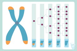
Researchers at the University of Wisconsin–Madison have described the way an enzyme and proteins interact to maintain the protective caps, called telomeres, at the end of chromosomes, a new insight into how a human cell preserves the integrity of its DNA through repeated cell division.
DNA replication is essential for perpetuating life as we know it, but many of the complexities of the process — how myriad biomolecules get where they need to go and interact over a series of intricately orchestrated steps — remain mysterious.
Every time a cell divides, the telomeres at the end of the long DNA molecule that makes up a single chromosome shorten slightly. Telomeres protect chromosomes like an aglet protects the end of a shoelace. Eventually, the telomeres are so short that vital genetic code on a chromosome is exposed and the cell, unable to function normally, enters a zombie state. Part of a cell’s routine maintenance includes preventing excessive shortening by replenishing this DNA using Polα-primase.
At the telomere construction site, Polα-primase first builds a short nucleic acid primer (called RNA) and then extends this primer with DNA (then called RNA-DNA primer). Scientists thought Polα-primase would need to alter its shape when it switches from RNA to DNA molecule synthesis. Lim’s lab found that Polα-primase makes the RNA-DNA primer at telomeres using a rigid scaffold with the help of another cog in the telomere replication machine, an accessory protein called CST. CST acts like a stop-and-go sign that halts the activity of other enzymes and brings Polα-primase to the construction site.
The researchers also got a glimpse into how Polα-primase might initiate DNA synthesis elsewhere along the length of a chromosome. Other scientists have also found the CST–pol-α-primase complex at sites where DNA damage is being repaired and where DNA replication has stalled.
The researchers built the structural model of CST–Polα-primase using an advanced imaging technique called cryo-electron microscopy single-particle analysis. In cryo-EM, rapidly frozen samples are suspended in a thin film of ice, then imaged with a transmission electron microscope, resulting high-resolution, 3D models of biomolecules like the enzymes at work in DNA replication.
Among these ideas are capturing how CST–pol-α/primase works in more detail. The researchers also want to map the entire human telomere replication process, and they’re studying how CST–pol-α/primase terminates its activity once the DNA at telomeres is copied.
University of Wisconsin-Madison
