AI and interpretive algorithms
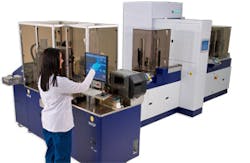
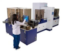
An innovative and collaborative approach to pre-analytics has resulted in original devices that have made microbiology collection processes simple and easy. Many of these collection and transport systems have been proven to advance the quality of traditional and contemporary microbiology assays. Flocked swabs and other liquid-based microbiology collection devices together with full laboratory automation (FLA) for microbiology, allow clinical laboratories to fully automate their culture testing. FLA includes specimen processing, smart incubation, digital imaging, and effective algorithms for automatic segregation of bacterial cultures, followed by automated colony selection and setting up of identification and susceptibility testing.
Collections devices and full laboratory automation
Automatic specimen processor units are a solution for preanalytical microbiology. These are open platforms, modular instruments, and can address all aspects of automated microbiology specimen processing: planting and streaking, Gram slide preparation and enrichment broth inoculation, and more. These systems can automate the workflow of the laboratory and allow the freedom to walk away from specimen set up and focus on higher level tasks. Most importantly, automated specimen processing is what microbiologists have asked for in their ideal laboratory.
Testing efficiencies, quality, and safety

FLA allows samples to be evaluated faster and without the need for additional staffing. The system allows for earlier growth detection as culture plates are in a continuous incubation situation at the correct temperature and atmosphere for optimal growth. There is no constant opening of the incubator doors to retrieve plates subjecting the cultures to suboptimal conditions, and plates are not left on the bench top for hours without proper incubation and atmospheric requirements. These efficiencies will allow for a new paradigm for what we think we know about incubation times. For example, traditionally urine cultures need to be incubated for 16-18 hours prior to the selection of colonies for further identification and susceptibility testing. With FLA and continuous incubation these cultures can be read as early as 10-12 hours. This new shift will occur with other types of specimens as we see improved bacterial growth of all cultures through defined, uninterrupted incubations. FLA allows for safe work up by laboratory staff without the need to be exposed to plates possibly growing highly infectious agents, and efficiencies will be gained by never needing to touch a negative plate again. The system also offers increased traceability using a bi-directional connection with the laboratory information system (LIS).
Artificial intelligence
Automated specimen processing and reading systems can be used to optimize laboratory resources for increased productivity. New AI/IA software uses artificial intelligence (AI) and interpretive algorithms (IA) to aid laboratory personnel with culture reading to allow for further optimization in the laboratory freeing up staff to concentrate on more difficult tasks.
AI/IA systems use a selection of highly sophisticated algorithms that will pre-assess, and pre-sort culture plates allowing microbiology laboratories to then read, interpret, and segregate bacterial cultures with the click of a button. As examples, algorithms that are currently on the market include:
- Segregation of chromogenic media for the detection of methicillin-resistant Staphylococcus aureus (MRSA);
- Digital analysis of chromogenic media for vancomycin-resistant Enterococcus (VRE) screens; and
- Pre-sorted digital segregation of urine culture quantitation.
Several currently available algorithms have been submitted to the Food and Drug Administration to allow for auto-verification and automatic release of results with no technologist intervention. Laboratories can also use the software to gather and analyze specimen results together with pertinent clinical patient information to optimize their algorithms. For example, algorithms can combine culture results with the sex and age of the patient to determine if small numbers of organisms detected in urine cultures need to be screened for group B streptococci.
In general terms, here’s how the software works: First a ‘time zero’ image is taken of the culture plate, then at user-defined incubation times additional images are taken and compared to the time zero image. Specifically, for chromogenic agars, the first question that is then asked is, “Is there something present now that was not present at time zero?” If the answer is yes, then the next question is, “What is the color of what is now present?” The final question that is asked is, “What hue is the color that is now present?” If the chromogenic media has growth, that growth is green, and the green colonies are the ‘right’ color (lime green for this particular media), then the AI tells you that the culture is presumptively positive. Conversely, if there is no growth or if the growth present is not the color and hue needed to be considered positive, then the AI will pre-sort that the culture as most likely negative. In addition, the software can determine colony counts of urine cultures and pre-sort these cultures into categories of:
- No growth;
- 100-1000 CFU/mL;
- 1000-10,000 CFU/mL;
- 10,000-100,000 CFU/mL; and
- > 100,000 CFU/mL with great accuracy.
Laboratory staff will also gain efficiencies as they will be able to focus on plates that AI/IA cannot yet deal with. In addition, using these algorithms there is not the variability that we see with human interpretation, i.e., the algorithms always make the same decision each time and never deviate from standard operating procedure.
Training and quality assurance
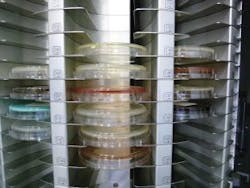
An extension of the above discussed advances of FLA, include an added advantage of utilizing the system for efficient training and quality assurance (QA) activities. Such activities would include utilizing images of known organisms and cultures for more efficient training of new staff members. Training technologists to recognize normal organism morphology could be expedited using the plethora of stored images of previous cultures. Technologists would be able to visualize multiple images taken over time on previous cultures along with the progression of culture results to learn how clinical microbiology decisions are made in the work up of various culture types. It could take months or longer for an isolate of Listeria monocytogenes, for example, to come into the laboratory, but with stored images along with Gram stain images, new staff can be trained to recognize these and other less commonly isolated organisms the first time they see it in a real time culture.
Laboratory QA is designed to detect, reduce and correct deficiencies and errors in laboratory practices to release quality patient results and involves all parts of testing: pre-analytical, analytical, and post-analytical processes. CLIA regulations (Subpart P) address specific quality assurance requirements and The Code of Federal Regulations (42 CFR 493) states laboratories “must establish and follow written policies and procedures for a comprehensive quality assurance program that is designed to monitor and evaluate the ongoing and overall quality of the total testing process.” Further it states that the QA program must:
- Assess the effectiveness of the laboratory’s policies and procedures;
- Identify and correct problems;
- Assure the accurate, reliable, and prompt reporting of test results; and
- Assure the adequacy and competency of the staff.1
Liquid based-microbiology (LBM) collection and transportation devices together with FLA can assist in a quality management system to consistently focus on meeting healthcare provider and patient requirements to enhance their satisfaction with the laboratory. LBM collection devices are manufactured to collect the best patient sample possible and maintain specimen integrity and organism viability until the specimen is processed in the laboratory resulting in superior patient results.
As just a few examples, automated specimen processing is a barcode-driven system that will reduces errors, improves accuracy of specimen processing, and eliminates transcription and transposition errors. Intuitive software and touch screen commands make all operations fast and simple, minimizing hands on time by the staff. FLA systems are flexible and can be designed to be modular and scalable to meet the unique needs of each unique laboratory regardless of space restrictions. Utilizing smart incubators and digital microbiology, users find that they are able to read plates more efficiently and accurately, detecting clinically relevant growth earlier than with traditional incubators and manual reading. This allows the laboratory to report more actionable results to clinicians faster to optimize patient care. In addition, images captured with digital microbiology can be used for blinded proficiency testing of laboratory personnel as part of a comprehensive competency assessment program. Lastly, new software AI/IA can accurately pre-assess growth on any manufacturer’s chromogenic agar for faster more accurate results, segregating presumptive positive cultures that may be missed on manual reading.2-4
In summary, today’s complete line of automated microbiology products leads the way, and will continue to do so with future innovations, to assist the laboratory with QA measures, staff training needs, and efficiencies for result reporting to improve patient outcomes.
REFERENCES
- American Academy of Family Physicians. CLIA and Quality Assurance. Quality Assurance, CLIA and Your Lab. https://www.aafp.org/practice-management/regulatory/clia/quality-assurance.html. Accessed Jan. 28, 2019.
- Faron ML, Buchan BW, Coon C, Liebregts T, et al. 2016. Automatic Digital Analysis of Chromogenic Media for Vancomycin-Resistant-Enterococcus Screens Using Copan WASPLab. Journal of Clinical Microbiology, 54(10), 2464-2469. doi:10.1128/jcm.01040-16.
- Faron ML, Buchan BW, Vismara C, Lacchini C, et al. 2016. Automated scoring of chromogenic media for detection of methicillin-resistant Staphylococcus aureus by use of WASPLab image analysis software. J Clin Microbiol 54:620 –624. doi:10.1128/JCM.02778-15.
- Kirn TJ. 2016. Automatic digital plate reading for surveillance cultures. J Clin Microbiol 54:2424-2426. doi:10.1128/JCM.01279-16.
About the Author

Susan E. Sharp, PhD, DABMM, FAAM
Ph.D., DABMM, FAAM, serves as Scientific Director for Copan Diagnostics, Inc., U.S. Dr. Sharp’s most prominent area of interest has centered on cost-effective, clinically-relevant, diagnostic microbiology. Sharp has served as a director for microbiology laboratory services for over 30 years.