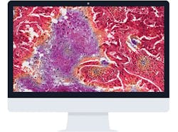
Transforming the practice of pathology with digital technology
Over the past three decades, the practice of pathology has evolved tremendously. In the 80s and 90s, viewing microscopic images was limited to looking through a pair of microscope oculars. Publications and presentations took hours of preparation using 35mm cameras that were adapted to the microscope. The exposed film was sent to a developer, and the resulting slides were submitted for journal publication or projected at a teaching conference on a standard carousel projector.
Some medical centers were fortunate enough to be able to invest in projection microscopes. A luxury for many, these “microscopes on wheels” could be transported from the laboratory to lecture hall to conference room. Images were projected directly from the slides to the screen. There was no video cable or computer, just a microscope, light source, and projection lenses.
Twenty-five years later, digital cameras transmit images from microscopes to remote computers for rapid assessment of frozen sections and fine needle aspiration preps in real time. “Whole slide imagers” are able to produce a high-quality digital image of a microscope slide within minutes which can be sent anywhere via the world-wide web for collaborative review. Whether for clinical, research, or educational applications, digital technology has transformed the way pathology is practiced today.
The pros and cons in a nutshell
What is driving the trend toward digital whole slide image technology? The biggest driver is the realization that being able to digitally capture an entire stained slide offers greater flexibility and utilization of microscopic images. Images can be annotated and shared easily among network users. New laboratory workflows reduce the handling of glass slides and improve pathologists’ flexibility and productivity. These new workflows also reduce the risk of loss of important patient material and reduce costs for managing slide archives.
Why isn’t everyone embracing digital whole slide image technology? Cost is a major factor. Although they’re trending down, investment costs for an image scanner and storage infrastructure can be formidable. In addition, support for implementation and operation of a digital pathology system requires teamwork across departments to ensure success. Pathologists who are unfamiliar with whole slide imaging technology may be overwhelmed and not want to depart from their familiar processes.
In fact, change is difficult, and it may require an investment of time, money, and trust before rewards are seen. To achieve success in this new practice of pathology, we must help our colleagues and ourselves overcome this resistance to change.
Process: decision-making strategy
In the last five years, whole slide scanners have improved in quality and come down substantially in cost. Larger medical centers find them invaluable in the preparation of material for research and teaching, and for support of many clinical applications such as quality assurance and tumor boards. Some of these centers are ready to take a bold next step by embracing telepathology to support their remote practice partners by collaborating on difficult cases. How will these centers change their current practice paradigm so they can realize the full potential of their digital pathology system?
A change of this magniude benefits by having a well-defined strategy that considers departmental and organizational goals. Knowing what needs to be achieved, when it needs to be achieved, and by whom it needs to be achieved provides a framework to guide purchasing decisions, timelines, and even staffing decisions.
There is much to be considered when purchasing a whole slide imager. Quality of images, magnification options, throughput, cost, and service are key factors in the decision-making process. Also to be considered is the relative value of each of these factors in case one needs to be compromised for another.
Other costs to be considered include those of an image storage server and of the technical staff to provide support for the service. Each whole slide image can range from 300 megabytes to several gigabytes. Adequate storage and backup of the image repository is critical to the long-term success of a digital pathology service. Staff support from both the laboratory and hospital information services is also required. These partners are vital to ensure the smooth operation of the digital pathology service from image acquisition to workflow management to the archiving of the images and their associated cases.
Implementation
Using a project management approach will assist with defining objectives, timelines, responsibilities, and key players to help ensure a successful process from purchase through implementation. The project will require the collaboration of a multidisciplinary team including organizational leadership, ancillary network experts, pathology staff, and, as needed, members from the vendor team.
The Team Leadership must provide a clear vision of what needs to be accomplished and when. It is their responsibility to articulate the project goals and to provide the resources, support, and commitment needed for success.
All team members must collaborate to consider hardware needs, calculate storage needs, and create a reasonable timeline to complete each phase of the project. It is critical to clarify the parameters of the project to prevent “scope creep.” Too often, implementation of a new work process can be sidetracked if time is spent on details not originally intended to be part of the project’s scope of work. Regular meetings to define tasks, assign responsibility, and report progress are vital to ensure the project stays on track.
Most team members will be juggling multiple priorities. Agendas, meeting notes, and updates to key stakeholders will assist in maintaining accountability for the work being done.
Key factors
Missteps can be avoided by focusing on a few specific considerations. Training, testing, standardization, and support are all key factors to ensure the successful implementation of a strong digital pathology service:
- Training: Do not underestimate the importance of proper training and support. Some staff may want to forego formal training, believing they can best learn by doing. Then, if the system does not perform as expected, they may place blame on product inadequacies instead of lack of user knowledge. Training and written documentation of processes for user reference are critical to success.
- Validation testing: Whole slide images must be validated to ensure that their quality is on par with glass slides, and the workflow platform must be validated for performance. Documentation of validation testing must be completed and maintained on file for regulatory inspections.
- Standardized workflows: Documentation of the new workflows is critical to ensure that all users and support staff know what steps happen and in what sequence. This documentation serves as a roadmap for how the workflows are navigated. At the technical level, work processes for scanning slides, accessioning cases, and triaging the work must be defined, and all staff must follow them. If work is standardized and sound quality control measures are in place, these activities become part of the lab’s daily routine to support their pathologists’ digital service.
- Pathologist support: Some pathologists may be reluctant to change to using whole slide images for quality assurance, tumor board activities, case reviews, or other routine work. If a standardized high quality digital service is provided by the laboratory, pathologists will begin to gain confidence in the new workflows. Users who become early adopters can support their colleagues as they come on board with this new way of work.
Pathology workflows
A repository of whole slide images does not constitute a digital pathology service. It is the work that can be accomplished with these images that provides the real value.
Most whole slide scanners are packaged with proprietary software that facilitates pathology workflows. Annotation and measurement tools can assist with research, preparation of study sets, and tumor board presentations. Quality assurance activities can be streamlined and documented for regulatory review. Collaborative case reviews between colleagues can be performed in real time whether the pathologist partner is down the hall or across the continent.
Third-party platforms that are scanner agnostic and can integrate with a user’s laboratory information system (LIS) have been proven to expand the utility of an organization’s repository of whole slide images. No longer are laboratories constrained by proprietary software specific to an individual scanner. Instead, pathologists and laboratories can take full advantage of the benefits offered by whole slide digital images regardless of the scanner that created them or the LIS storing the patient’s information.
These powerful new platforms contain the tools that elevate a digital pathology system to a truly comprehensive clinical workflow suite.


