The times they are a-changin’ in clinical microbiology
Earning CEUs:
Laboratories have been practicing clinical microbiology the same way for decades, however over the last several years things have started to change; change is necessary as microbiology faces big challenges. We are seeing an increased workload due to screening and diagnosing agents of hospital-acquired infections, multiple drug resistant organisms, and are facing the pressure of producing faster results to be able to discharge patients from the hospital more quickly. And we are doing all of this, and more, while facing decreasing resources. We are receiving less reimbursement for the testing we perform, and we are undergoing laboratory consolidations without attaining the workforce necessary to perform the additional testing—which places added pressure on our already stretched staffing levels. All of this is in the face of a shrinking workforce due to retirement (20 percent of clinical microbiologists will retire in the next 5 years1) and the lack of interest of people entering the field. We must find efficiencies for the future.
Our colleagues in clinical chemistry, hematology, and immunology perform much of their work on large automated lines containing instrument after instrument, which perform patient testing 24/7 and report a vast majority of results with no intervention by laboratory staff. Of course, our colleagues have the advantage of having their specimens largely submitted in blood tubes of all the same size, which makes automation possible. In microbiology, on the other hand, we handle a multitude of specimen types with many varying consistencies making automation more difficult. But advances in specimen collection devices for clinical microbiology have made automation a reality. The introduction of the flocked swab in 2003 allowed throat, wound, genital, etc. collections to be liquid-based with over 90 percent of the specimen collected being delivered to the medium in the tube, as opposed to less than 35 percent recovery utilizing traditional swabs. Flocked swabs can be used not only for the collection of specimens for the recovery of aerobic, anaerobic, and fastidious bacteria but have also been shown to be excellent collection devices for yeasts and molds,2 as well as to be useful for molecular assays.3 Additionally, with the advent of other new innovative collection devices for urine, sputum, and feces, these specimen types can now be collected into a similar sized tube and be ready for placement onto automated processing platforms.4
Liquid based microbiology: the key to simplifying collection, transport, and processing of samples
Collection and transport systems consisting of a flocked swab and a tube with 1mL of Amies liquid media in which to elute the specimen can be used for multiple types of testing. One study looking for the presence of Fusobacterium necrophorum in children presenting with pharyngitis was able to perform five culture plates, one rapid point-of-care assay, and two molecular assays from the same specimen collection.5 This ensures quality and standardization for all testing performed. Another study found that liquid-based microbiology not only allows for specimen collection optimization, but helps to reduce cost due to the smaller number of different devices that need to be stored, reduces the time spent by medical and nursing staff as there is less confusion regarding collection device selection (one flocked swab type for all testing), and there were fewer samples being collected from the patient using multiple different devices. This last advantage relates to patient satisfaction and time saved for laboratory staff, as there are fewer samples to handle per patient.6 Chapin and colleagues found that the use of rectal flocked swabs have the advantage of being able to provide actionable results sooner in a pediatric emergency room population when using molecular testing for stool pathogens as compared to routine stools which can be difficult to collect in this population.7
These advances in liquid-based microbiology specimen collection devices have opened the door to automated specimen processing in our laboratories, which has been shown to provide increased recovery of pathogenic organisms. A study by Michnik and colleagues using stool specimens showed better recovery of microorganisms versus manual inoculation, and that the quality of colony growth and isolation was superior to that of manually streaked plates. In particular, they were able to show that the number of Campylobacter organisms isolated from stool cultures increased with automated specimen processing as compared to manual processing.8,9 And increases in the number of Group B streptococci isolated from pregnancy screens can be realized when using flocked swabs and automated processing, as compared to routine swabs and manual processing.10
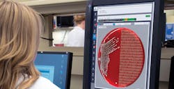
Full laboratory automation, digital microbiology, and artificial intelligence
Liquid-based microbiology specimen collection devices opened the door to automated specimen processing, which has in turn opened the door to full microbiology laboratory automation in our laboratories. Full laboratory automation ensures that cultures are at a constant temperature with appropriate atmospheric conditions for optimal growth. Images are taken of plates and decisions on further work are made by the microbiologist after reviewing these images. Without the need to constantly take culture plates out of the incubators for reading (and consequently leaving them on the bench for hours where bacteria are not growing optimally), organisms will grow faster allowing for more rapid work up of pathogens and the ability to provide actionable results sooner to our clinicians. And now that full laboratory automation has digitized our reading of cultures, artificial intelligence (AI) will allow innovative algorithms and specialized software to automatically report culture results, similar to our core-laboratory colleagues. AI in the clinical microbiology laboratory amplifies human ingenuity and is the next frontier in full laboratory automation as it focuses on algorithms to automatically read and interpret growth on plates, count colonies, and recognize colony morphology.
The essential algorithms for AI for microbiology can be grouped into four categories: 1) chromogenic detection which automatically detects color of colonies on chromogenic media plates; 2) colony counting with growth/no growth discrimination which quickly screens negative plates and segregates no growth and no significant growth plate from those with growth; 3) phenotypic colony recognition which examines colonies on non-chromogenic plates, comparing them against a library of thousands of colony images to mate the phenotypic characteristics and assign predictive values for identification; and 4) application of user-defined expert rules to filter outputs and reporting.
AI vs. manual reading
Chromogenic algorithms have reported to work with 100 percent sensitivity in two large multi-center publications using multiple types of chromogenic agar for screening of pathogenic organisms.11 One hundred percent sensitivity means that the software does not report any culture as negative that manual reading would have called positive. Similar results have been shown with screening cultures for Streptococcus pyogenes from throat specimens and with vaginal/rectal pregnancy screens for Group B streptococci; again, in these studies the software did not report any culture as negative that manual reading called positive.12 Indeed, the software was able to find positive cultures that were missed by manual screening. Additionally, the vancomycin-resistant enterococci (VRE) publication referred to previously was also evaluated to determine the cost saving that would be gained using the algorithm pre-assessment as opposed to manual microbiology processes. They were able to show that with the almost 88,000 negative VRE screens included in their study, approximately $450,000 in labor costs could have been saved through the use of automation versus manual techniques.
Labor savings and turnaround time reduction
Additionally, algorithms are available that will determine not only growth/no growth from urine cultures (our highest volume specimen in clinical microbiology), but also use segregation software along with chromogenic agars to sort these cultures into user-defined groups of insignificant growth, mixed growth, and those that are of significant counts that would need identifications and susceptibilities performed. Preliminary investigations show that when using imaging and segregation software you can automatically categorize over 70 percent of urine culture results for batch release or additional testing leaving a minority that would need to be reviewed manually by staff. The software was shown to decrease the turnaroud time for culture results, improve workflow and quality, and allow for cost savings as laboratories were able to reduce their need to hire new employees—despite an increase in retirements and increases in workload. AI software eliminates mundane tasks for staff, allowing them to concentrate their efforts on the more difficult cultures and laboratory testing. A study from Faron and colleagues looked at over 3,000 urine cultures plated to chromogenic agar and showed that upon secondary review of originally discrepant results, AI software showed a sensitivity of 99.8 percent in the determination of positive (> 10,000 CFU/mL) and negative (< 10,000 CFU/mL) urine cultures with a specificity of 86.2 percent. This study also showed a reduction in turnaround time of six hours with negative cultures and of eight hours with positive cultures.13
AI also has a role in assisting in the interpretation of growth from traditional media (non-chromogenic agars). A recent study from Milwaukee, WI evaluated the ability of AI software to determine colony counts from urine cultures using routine blood and MacConkey agars. Preliminary results on over 5,000 urine cultures showed agreement of AI software in the determination of colony counts with manual reading at a sensitivity of 99 percent for significant cultures (> 10,000 CFU/mL), and 92.6 percent for insignificant cultures (< 10,000 CFU/mL). Additional samples will be added to this study for a total of over 15,000 urine cultures looking at the ability of AI software to differentiate colonies on blood and MacConkey agars compared to manual analysis.14
Colony counts
Additional investigations have looked at the ability of the AI software to not only determine colony counts, but also its capability to identify mixed cultures using traditional urine culture media. Preliminary data suggests that colony count software determinations can very accurately be made and highly agree with manual counts (R2value = 0.9152). In addition, they found that agreement of AI software with manual reads of cultures containing two morphologies of organisms is 97.4 percent and reaches 95.0 percent with cultures containing three or four different morphotypes of organisms. These investigators also looked at the ability of AI software to determine clinical cutoff (<10,000 CFU/mL) and to discern the numbers of isolates present from urine cultures, with ≥ 3 morphologies indicating contamination. After review of over 5,000 urine specimens plated to routine blood and MacConkey agars there was 92 percent agreement between the software and manual reading of the cultures for those that needed identification and susceptibilities performed, 96 percent agreement for those that showed contamination, and 99.6 percent agreement for no growth results.15,16
AI and rainbow unicorns
Investigators have shown a reduction of approximately 50 percent in the time necessary to perform specific tasks in the laboratory when using full laboratory automation and AI algorithms. For example, when assessing the tasks of urine screening and reading, picking of colonies for further analysis, and screening of MRSA cultures, it was shown that these three tasks took approximately 17 hours daily before the implementation of full laboratory automation and AI algorithms; after implementation these same three tasks took approximately nine hours per day. Multiple laboratories have been able to show that post implementation of full laboratory automation with and without AI algorithms, increased workload can be handled with decreased staffing, adding weight to the adage of “doing more with less.”
As vacancies and the demand for overtime continue to put stress on our remaining staff, microbiology laboratory automation and innovative reading algorithms can help to fill these vacancies by relieving workload, decreasing errors, and reducing the repetitive motion injuries associated with specimen processing and culture work up/reporting. We know that culture work can be a slow process, especially when compared to newer molecular techniques, but laboratory automation and associated improvements can allow for more rapid reporting of culture results. We also know that experienced clinical microbiologists are a rare breed (as rare as rainbow unicorns) and that to train new employees in microbiology can take months to a year. Automation in clinical microbiology will enable the redistribution of labor and allow microbiologists to focus on more complex tasks that need their attention.
In summary, liquid-based microbiology specimen collection devices, automated specimen processing, and digital imaging with AI reading algorithms are adding value to our microbiology laboratories now and will do so into the future as we work together to conquer the challenges we face.
REFERENCES
- Garcia E, Kundu I, Ali A, Soles R. The American Society for Clinical Pathology’s 2016-2017 vacancy Survey of medical laboratories in the United States. Am J Clin Pathol. 2018;149:387-400.
- J Clin Microbiol. 2014 Jun;52(6):2166-8.
- Gandhi B, Summerbell R, Mazzulli T. Evaluation of the Copan ESwab transport system for viability of pathogenic fungi by use of a modification of Clinical and Laboratory Standards Institute document M40-A2. J Clin Microbiol. 2018;56(2):e01481-17. Published 2018 Jan 24. doi:10.1128/JCM.01481-17.
- Chapin K, Duffy S, LeBlanc L, Bushey E, Stockmann C, Daly JA, Selvarangan R, Kanwar N, Jackson J, Pavia AT. Comparison of results obtained with the Film Array GI panel using recta-swabs and Carey-Blair stool from patient with gastroenteritis in a pediatric emergency department. Poster Presented at the 2016 Clinical Virology Symposium, Daytona, FL.
- Fontana C, Favaro M, Favalli C. How liquid based microbiology can change the workflow in the microbiology laboratories. Advances in Microbiology, 2013, 3, 504-510.
- Van TT, Cox LM, Cox ME, Dien Bard J. Prevalence of Fusobacterium necrophorum in dhildren presenting with pharyngitis. J Clin Microbiol. 2017 Apr;55(4):1147-1153. doi: 10.1128/JCM.02174-16. Epub 2017 Jan 25.
- Mischnik A, Trampe M, Zimmermann S. Evaluation of the impact of automated specimen inoculation, using Previ Isola, on the quality of and technical time for stool cultures. Ann Lab Med. 2014;35(1):82-8.
- Baker J, Hermann A, Bitting C, Culbreath K. Improved Isolation of Bacteria in Stool Cultures Using the COPAN FecalSwab Processed by COPAN WASP Compared to Manual Plating Methods. Poster presented 2017 Microbe Meeting, New Orleans, LA.
- Buchan BW, Olson WJ, Mackey TL, Ledeboer NA. Clinical evaluation of the walk-away specimen processor and ESwab for recovery of Streptococcus agalactiae isolates in prenatal screening specimens. J. Clin. Microbiol. 2014, 52(6):2166. DOI: 10.1128/JCM.00374-14.
- Faron ML, Buchan BW, Coon C, et al. Automatic digital analysis of chromogenic media for vancomycin-resistant Enterococcus screens using Copan WASPLab. J Clin Microbiol. 2016; 54:2464-2469.
- Faron ML, Buchan BW, Coon C, et al. Automatic scoring of chromogenic media for detection of methicillin-resistant Staphylococcus aureus by use of WASPLab image analysis software. J Clin Microbiol. 2016; 54:620-6243.
- Dien Bard J, Nelson J, Mata K, Thrope D, Novak-Weekley S. Digital detection of group A Streptococcus using Colorex Strep A CHROMagar and WASPLab chromogenic detection module. Poster presented at 2018 28th ECCMID Meeting, Madrid, Spain.
- Pedna MF, Fantini M, Sambri V. Validation of image analysis software for automatic reading of Streptococcus agalactiae (GBS) isolates in prenatal screening specimens. Poster presented at 2018 28th ECCMID Meeting, Madrid, Spain.
- Faron, ML, Buchan BW, Relich, RF, et al. Digital image analysis to interpret urine cultures on blood and MacConkey afar. Poster presented at 2017 Microbe Meeting, New Orleans, LA.
- Faron, ML, BW Buchan, NA Ledeboer. Automatic Urine Culture Analysis Using CPSe Agar and the WASPLab Chromogen Detection Module. Poster presented at 2017 27th ECCMID Meeting, Amsterdam, Netherlands.
- Timm K and K Culbreath. Expert image analysis by COPAN WASPLab to evaluate urine cultures. Poster presented at 2017 Microbe Meeting, New Orleans, LA.
About the Author

Susan E. Sharp, PhD, DABMM, FAAM
Ph.D., DABMM, FAAM, serves as Scientific Director for Copan Diagnostics, Inc., U.S. Dr. Sharp’s most prominent area of interest has centered on cost-effective, clinically-relevant, diagnostic microbiology. Sharp has served as a director for microbiology laboratory services for over 30 years.