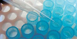
Slide makers and stainers: automating in the Hematology lab to improve efficiency
“Is this neutrophil hypogranular, or is my stain on the blink again? This can be the difference between catching myelodysplastic syndrome or not….”
“The sample shows thrombocytopenia for a 71-year-old male. Should this go to the pathologist?”
“The previous patient slide had Döhle bodies, but they were also hard to read. It’s a STAT but I’ll have to stain another slide, just in case….”
Have you ever found yourself dealing with these issues, or others like them?
The language of morphology
Reading slides at the microscope can pose many challenges with potentially perilous outcomes. Identification of cell abnormalities—such as the large azurophilic granules in toxic granulation, asynchronous maturation, micromegakaryocyte identification or quantitating variant lymphs—still requires a skilled human eye. Color, intensity, and contrast can be essential to differentiate white cells and aid in red cell and platelet characterization. Cells speak the language of morphology, and a skilled laboratorian translates this dialect into clinically actionable information. A poorly prepared slide can inhibit proper interpretation and potentially lose the opportunity to catch a disease in a timely manner.
Proper slide preparation involves not only adequate staining but also consistent cell distribution to facilitate interpretation at the scope. Struggling to identify the monolayer only adds to the time-crunch of the morning run. What about a low WBC count? Should we extrapolate from the 28 cells we identified, or take the time to make more slides? It has been documented that even a 100-cell count may have insufficient precision. Varying situations may also call for additional slides—for example, in teaching facilities where evaluation is done by multiple residents, or when oncologists and clinicians request to review patient slides. Fortunately, modern slide-making and staining technology provides greater workflow efficiency and stain reliability.
The art of slide prep
As laboratories take steps toward automating the slide- making and -staining process, there are some important aspects to consider. Trained laboratorians know that in addition to the skill of reading cells, there is also an art to slide preparation. Standardization is essential to an effective slide-making process. When I studied clinical laboratory science, I was told by an instructor that it would take a whole box of slides before I would learn to smear correctly. Eventually, I perfected the skill and learned to slowly mix the tube by inverting it and observing how the blood grabs the tube walls. This told me how quickly to pull the blood across the slide. Viscosity mattered: if it was thick, I learned to pull quickly; if it was thin, to pull slowly. The angle needed to be adjusted accordingly as well. If the WBC count were low or I was working with a multi-headed scope, multiple slides might be needed. Like most of my readers, I learned by doing.
When you as a lab leader are deciding whether to fully automate the slide-making and -staining process, the quality of the smear is of utmost importance. Upon installation of the automated system, vendors will train laboratorians and help establish stain preferences on the newly installed slide maker/stainer (SMS). There is usually wide variability in what is considered desirable in a specific laboratory, so ensuring there is a formal standard for your laboratory will help ease the installation process. Once the initial setup is performed, your vendor should support and empower your laboratory to make changes effortlessly, without a service call, as often as possible. Changes in support reagents, such as water supply or methanol, can make subtle differences, and you want to have the ability to adjust on the spot. Automating the staining process helps to insure the freshest stain, dependent on time or usage, and thus to deliver your protocol’s best performance. Having the same manufacturer for both instrumentation and stain reagents improves harmonization of the color with the process, along with vendor support.
Mimicking human skill
SMS instrumentation methods can vary in smear preparation technology, but slide consistency is part of the expectation for perfect slide-preparation, regardless of blood viscosity challenges. In many cases, very watery blood leads to inconsistent smear lengths or patterns and ends up requiring a manual smear preparation. However, today’s advanced technology can mimic human technique and create a perfect slide, even with very thin blood. This technology functions much like a laboratorian who observes the patient’s blood cling to the walls of a tube. It measures the blood residue, or cling of the blood, as it travels. The instrument then adjusts the slide-over-slide speed, much as a laboratorian would when completing this process by hand. This capability leads to reproducible smears regardless of variation in blood consistency, making the monolayer of a slide easier and quicker to find for the best workflow.
Automating the slide-making and -staining process also helps the laboratory overcome clerical errors and time delays. Typically, a laboratory with more than 40 slides per day can benefit from automation. The instrument will use information from the laboratory information system (LIS) for label making, helping to eliminate misidentification. Selecting an instrument with a sample-quality alert process will enable technologists to quickly address aspiration errors (e.g., clots which can affect cell distribution). Additionally, instruments are able to send alerts when a sample is skipped or dropped. And, the ability to follow the slide and its estimated completion time allows the staff to multitask more efficiently.
The hands-off process is most effective when the SMS is connected to the CBC analyzer, as decision-making without intervention allows for a true automated process. This can be achieved with a CBC analyzer that will communicate with the SMS beyond triggering a slide or not. Rules based on the CBC results will overcome the workflow exceptions and provide expanded benefits. Additionally, rules can be set to trigger additional slides based on specific information—demographics, flagging, the patient’s physician, department, CBC, etc.—much as the laboratorian would do. The laboratorian, or an automated cell counter, can then add the counts from the additional slides for maximum number of cells included in a diff. Expanded rules help deliver better patient care without extra effort.
Toward a greener lab
The environment will also benefit from a vigilant instrument pre-sale investigation. Some states enforce a zero-tolerance policy with random testing for mercury and heavy metal waste down drains. Lab leaders can ask the vendor for an effluent waste letter certifying a profile of elements for which they tested. An environmental officer can use this certificate to evaluate local and EPA limits. Modern instrumentation can split the waste and allow friendly material to drain while collecting the hazardous metal and methanol waste separately for proper disposal. This helps lower disposal costs and, more importantly, limits environmental impact.
It is imperative that laboratories’ diagnostic tools are optimal for interpreting and reporting even subtle changes in cells with confidence. Automating slide-making and -staining should not compromise quality, but rather add efficiency and standardization. This is achieved by the instrument’s ability to adapt to blood sample variability, easily optimize stain protocol for best color quality, and make slides based on clinical or workflow needs with extended rule writing—all while limiting environmental impact. Instruments available on the market today vary considerably in the features they offer, and you want to ensure your laboratory chooses a solution which serves as many of its needs as possible.