Individualized risk prediction in Barrett’s esophagus
Barrett’s esophagus (BE), which is a precursor to esophageal adenocarcinoma (EAC), is an increasing healthcare challenge in the United States, but new tools are quickly emerging to combat the uncertainty associated with the disease. Barrett’s esophagus management decisions are often based on subjective diagnoses, so better diagnostic and prognostic techniques are needed to augment BE patient care. Here, we summarize a number of tools available to clinical decision-makers to aid diagnoses and predict the risk of disease progression, which will optimize patient surveillance intervals and guide therapeutic interventions.
The clinical problem
There are an estimated 12 million to 17 million people who have BE in the United States.1 Although malignant progression is rare in BE, a small subset of patients will develop EAC,2,3 which has a five-year survival rate of less than 20 percent.4 Furthermore, the incidence of EAC continues to rise in the U.S. Treatment options for EAC are limited, so early detection is critical for optimal patient management.
Currently, patients with BE are surveilled by endoscopic biopsies with the goal of detecting disease progression early. Diagnoses are not always clear-cut, and can be inconclusive even after review by a specialized pathologist.5 Recent guidelines published by the American College of Gastroenterology recommend endoscopic ablative therapy for patients diagnosed with high-grade dysplasia, but there is uncertainty about the surveillance interval, true risk of progression, and treatment recommendations for patients with low-grade dysplasia or who are indefinite for dysplasia, and for patients with non-dysplastic BE.6 Therefore, it can be difficult to distinguish patients with BE who are at high risk for progression to EAC from those whose disease will not progress, thus making surveillance and treatment decisions challenging.
Enhanced diagnostic techniques
Diagnosis of BE relies on endoscopic recognition of salmon-pink colored esophageal lining and confirmation of the presence of columnar epithelium in pinch biopsies taken during endoscopy. While this approach is valuable, it is limited by the random nature of the sampling and observer variability in the histologic diagnosis. New sampling techniques have been developed that aim to overcome some of these limitations. One such technique is computer-assisted brush biopsy, which can be used as an adjunct to standard biopsy to detect BE and to aid in the identification of dysplasia.7,8 Brush biopsies allow for more esophageal tissue to be sampled, thus improving tissue coverage and increasing the potential to detect BE and dysplasia in a single endoscopy.
Volumetric laser endomicroscopy (VLE) is an endoscopic technique that utilizes advanced imaging technology to generate three-dimensional images of tissue in vivo.9,10 Compared to other in vivo imaging methods, VLE increases imaging depth and decreases acquisition time and can be used to guide biopsy samples to locations that potentially have abnormalities, and to mark regions for therapeutic intervention. In addition, non-endoscopic tissue collection devices have been developed as a minimally invasive option to increase patient compliance and increase detection rates.11,12 The collection device is swallowed, and then it collects cells as it is removed back out of the mouth of the patient. Importantly, non-endoscopic devices have the potential to identify patients with BE that might have been overlooked if they were initially unwilling to undergo standard endoscopy.
New risk prediction approaches
While timely identification of patients with BE is an essential first step, subsequent monitoring and treatment recommendations are not always clearly defined. Clinical and pathologic variables are inadequate to predict which patients will progress to EAC, and over-surveillance of patients is common due to uncertainty in the diagnostic stage and anxiety relating to the unknown risk of developing EAC.13 Healthcare providers require tools to accurately stratify patients based on their risk of disease progression. Such tools will allow increased surveillance and early therapeutic intervention for the subset of patients at high risk for progression to EAC, and permit longer surveillance intervals for patients at very low risk.
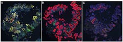
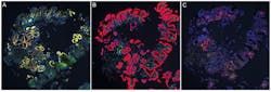
Several approaches have been developed for risk stratification in BE. One approach, which has been validated in clinical studies and is commercially available as a laboratory-developed test (LDT), is a tissue systems pathology assay that quantifies multiple key biomarkers in BE biopsies to produce an individualized risk score for progression.14 The technology underlying this assay quantifies not just epithelial abnormalities that are indicative of progression, but also stromal changes, such as angiogenesis and infiltration of specific immune cell subsets that play important roles in tumor development and progression.15 This approach utilizes immunofluorescence labeling to detect a series of biomarkers on formalin-fixed paraffin-embedded (FFPE) tissue sections (Figure 1). After capturing whole slide digital images of the tissue, specialized image analysis software automatically segments specific subcellular compartments and tissue structural components and quantifies biomarker expression patterns in the context of the cellular and tissue architecture. This imaging approach has the advantage of assessing multiple key cell types, including immune cells, and multiple pathways of malignant progression, while maintaining essential spatial and contextual information. A multivariable classifier is then used to integrate the quantitative biomarker and morphology data into a risk score, which is used to estimate the individual patient’s risk of disease progression within the next five years, as well as to identify patients who might already have prevalent HGD or EAC.14,16
Other approaches have assessed individual biomarkers or panels of biomarkers stained by immunohistochemistry on FFPE tissue and found a modest benefit for risk prediction.17,18 However, these tools rely on manual interpretation of biomarkers on tissue slides and have not yet been implemented for risk prediction in clinical practice. Other approaches have examined mutations using next generation sequencing and PCR and found that patients who progressed to HGD or EAC had an increased mutational load.19,20 While not currently commercially available for clinical testing, these methods have the advantage of high-throughput detection of multiple mutations that may indicate future malignant progression. The drawback of these methods is the loss of spatial context of molecular changes. Furthermore, these methods do not assess cellular changes in the stroma and morphologic changes that indicate risk of progression.
Individualized risk prediction methods will ease the unnecessary concern of patients whose BE is at low risk of progressing while highlighting patients at high risk to ensure they receive more aggressive care. Another important benefit is the potential for cost savings by extending endoscopic surveillance intervals in low-risk patients, and by early therapeutic intervention in patients at high risk of progression, which is expected to reduce the significant cost burden associated with cancer treatment and end-of-life care.21,22
New tools and new optimism
In summary, accurate risk prediction provides an opportunity to improve patient management by providing better outcomes while improving the efficiency of healthcare spending in the management of BE. Ultimately, the goal is to reduce the incidence and mortality of EAC in patients with BE, which can be accomplished by improving methods of early detection and intervention. The new tools that improve diagnostic accuracy and provide accurate risk stratification are an exciting step forward in this process.
REFERENCES
- Hayeck TJ, Kong CY, Spechler SJ, Gazelle GS, Hur C. The prevalence of Barrett’s esophagus in the US: estimates from a simulation model confirmed by SEER data.
Dis Esophagus. 2010;23(6):451-457. - Wani S, Falk G, Hall M, et al. Patients with nondysplastic Barrett’s esophagus have low risks for developing dysplasia or esophageal adenocarcinoma. Clin Gastroenterol Hepatol. 2011;9(3):220-227; quiz e226.
- Thota PN, Lee HJ, Goldblum JR, et al. Risk stratification of patients with barrett’s esophagus and low-grade dysplasia or indefinite for dysplasia. Clin Gastroenterol
Hepatol. 2014;13(3):459-465 e451. - Cancer Facts & Figures 2016 American Cancer Society.
- Yantiss RK. Diagnostic challenges in the pathologic evaluation of Barrett
esophagus. Arch Pathol Lab Med. 2010;134(11):1589-1600. - Shaheen NJ, Falk GW, Iyer PG, Gerson LB. ACG Clinical Guideline: Diagnosis and Management of Barrett’s Esophagus. Am J Gastroenterol. 2015;111(1):30-50.
- Johanson JF, Frakes J, Eisen D. Computer-assisted analysis of abrasive transepithelial brush biopsies increases the effectiveness of esophageal screening: a multicenter prospective clinical trial by the EndoCDx Collaborative Group. Dig Dis Sci. 2010;56(3):767-772.
- Anandasabapathy S, Sontag S, Graham DY, et al. Computer-assisted brush-biopsy analysis for the detection of dysplasia in a high-risk Barrett’s esophagus surveillance population. Dig Dis Sci. 2010;56(3):761-766.
- Swager A, Boerwinkel DF, de Bruin DM, et al. Volumetric laser endomicroscopy in Barrett’s esophagus: a feasibility study on histological correlation. Dis Esophagus. May 8 2015, doi: 10.1111/dote.12371.
- Leggett CL, Gorospe EC, Chan DK, et al. Comparative diagnostic performance of volumetric laser endomicroscopy and confocal laser endomicroscopy in the detection of dysplasia associated with Barrett’s esophagus. Gastrointest Endosc. 2016;83(5):880-888 e882.
- Ross-Innes CS, Debiram-Beecham I, O’Donovan M, et al. Evaluation of a minimally invasive cell sampling device coupled with assessment of trefoil factor 3 expression for diagnosing Barrett’s esophagus: a multi-center case-control study. PLoS Med. 2015;12(1):e1001780.
- Iyer P, Johnson ML, Lansing R, et al. Discovery, Validation and Feasibility Testing of Highly Discriminant DNA Methylation Markers for Detection of Barrett’s Esophagus Using a Capsule Sponge Device. Gastroenterology. 2016;150(4):S66-67.
- Cai JX, Campbell EJ, Richter JM. Concordance of Outpatient Esophagogastroduodenoscopy of the Upper Gastrointestinal Tract With Evidence-Based Guidelines. JAMA Intern Med. 2015;175(9):1563-1564.
- Critchley-Thorne RJ, Duits LC, Prichard JW, et al. A Tissue Systems Pathology Assay for High-Risk Barrett’s Esophagus. Cancer Epidemiol Biomarkers Prev. 2016;25(6):958-968.
- Prichard JW, Davison JM, Campbell BB, et al. TissueCypher: A Systems Biology Approach to Anatomic Pathology. Journal of Pathology Informatics. 2015;6:48.
- Critchley-Thorne RJ, Davison JM, Prichard JW, et al. Sa1257 A Tissue Systems Pathology Test Detects a Field Effect Associated With High Grade Dysplasia and Esophageal Cancer in Barrett’s Esophagus Patients. Gastroenterology. 2016;150(4):S259.
- Bird-Lieberman EL, Dunn JM, Coleman HG, et al. Population-based study reveals new risk-stratification biomarker panel for Barrett’s esophagus. Gastroenterology. 2012;143(4):927-935 e923.
- Horvath B, Singh P, Xie H, Thota PN, Sun X, Liu X. Expression of p53 predicts risk of prevalent and incident advanced neoplasia in patients with Barrett’s esophagus and epithelial changes indefinite for dysplasia. Gastroenterol Rep (Oxf). Oct 19 2015.
- Del Portillo A, Lagana SM, Yao Y, et al. Evaluation of Mutational Testing of Preneoplastic Barrett’s Mucosa by Next-Generation Sequencing of Formalin-Fixed, Paraffin-Embedded Endoscopic Samples for Detection of Concurrent Dysplasia and Adenocarcinoma in Barrett’s Esophagus. J Mol Diagn. 2015;17(4):412-419.
- Eluri S, Brugge WR, Daglilar ES, et al. The Presence of Genetic Mutations at Key Loci Predicts Progression to Esophageal Adenocarcinoma in Barrett’s Esophagus. Am J Gastroenterol. 2015;110(6):828-834.
- Gordon LG, Mayne GC, Hirst NG, Bright T, Whiteman DC, Watson DI. Cost-effectiveness of endoscopic surveillance of non-dysplastic Barrett’s esophagus. Gastrointest Endosc. 2014;79(2):242-256 e246.
- Hao J, Snyder SR, Pitcavage JM, Critchley-Thorne RJ. A Cost-Effectiveness Analysis of A Test That Predicts Risk of Malignant Progression In Barrett’s Esophagus. Value in Health. 2016;19(3):A6-7.
Aaron D. DeWard, PhD, serves as Research Scientist for Cernostics, Inc., provider of the TissueCypher Barrett’s esophagus assay.
Rebecca Critchley-Thorne, PhD, serves as Vice President, Research and Development, and co-founder of Cernostics, Inc.