Urine eosinophil method, no reagent expiration date, and shoe safety


Urine eosinophil method
Q: I am interested in a current procedure for evaluating urine eosinophils. I am especially interested in time stability for the specimens and comparison of Wright stain vs. Hansel stain. Could you please send me some information or direct me to a current source?
A: Eosinophils are seen in the urine in a variety of conditions, probably most importantly being drug-induced acute interstitial nephritis (AIN). In this case, the resulting renal dysfunction may be rapidly and effectively treated by discontinuation of the offending drug. Unfortunately, eosinophiluria is not specific for AIN, nor is it seen in all cases. Other conditions which show eosinophiluria include eosinophilic cystitis,5 atheroembolic renal disease,9 and schistosomiasis.4 Other diseases which have been found to be associated with eosinophiluria include rapidly progressive glomerulonephritis, acute prostatitis, postinfectious glomerulonephritis, and acute cystitis.2,6,7
If your urinalysis area does not have a cytocentrifuge, you may wish to refer specimens to another area which does, such as hematology or cytology. If no cytocentrifuge is available, slides can be prepared by placing a drop of concentrated urine sediment on a glass microscope slide and allowing it to air dry.
It is important that the specimen be fresh, if eosinophils are to be recognized.2 Slides should be prepared as soon as possible after voiding, certainly within three hours and preferably within one hour. The cells will rapidly disintegrate in urine and be unrecognizable.
Although it would be theoretically advantageous to identify eosinophiluria by flow cytometry, we are unaware of any such method, nor are we aware of methodology using blood cell counters.
Methodology:
Specimen preparation:
- Centrifuge 12 mL fresh, well mixed urine at 450 rpm for five minutes. Pour off 11 mL of supernatent urine. Re-suspend remaining sediment in 1 mL urine (12:1 concentration).
Slide preparation:
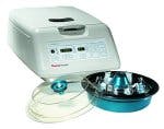
- Prepare slides using the Cytospin cytocentrifuge (Thermo Shandon Inc., Pittsburgh), according to manufacturers directions, as follows:
- Assemble Cytospin chambers, with glass microscope slide and Cytofunnel as directed. Prepare two slides for each specimen.
- Place well-mixed, concentrated sediment into the Cytospin sample chamber. Make sure that the fluid goes into the bottom of the funnel. The number of drops of concentrated sediment needed is based on the number of white cells present per high power (40x) field.
- If the urine protein is negative or trace, add one drop of 22 percent Bovine albumin to the chamber so that cells will adhere to the slide.
- Place Cytospin sample chambers into the Cytospin head, seal the head, place in the centrifuge, close the cover, and spin at 700 rpm for 10 minutes.
- Remove slides from chambers and allow to air dry.
- Alternatively when there is no cytocentrifuge available:
- Place one drop of concentrated urine sediment on a glass microscope.
- Spread slightly.
- Allow to air dry.
- Stain.
Staining:
- Stain with Hansel stain (Lide Laboratories, Florissant, MO), according to manufacturer directions, as follows:
- Dehydrate with methyl alcohol.
- Flood with Hansel stain; let stand 20 seconds to 30 seconds.
- Add distilled water; wait 20 seconds to 30 seconds.
Pour off and rinse with distilled water.
- Rinse with methyl alcohol. Re-rinse if necessary.
Examine the stained slides with low power (10x objective) and then oil immersion (100x objective). Report the percentage of eosinophils seen in 300 white blood cells. If there are less than 300 WBCs on the slide, report the percentage of eosinophils seen in the cells present.
Karen M. Ringsrud, MT(ASCP)
Assistant Professor
Dept. of Laboratory Medicine and Pathology
University of Minnesota Medical School
Minneapolis
References:
1. Corwin HL, Bray RA, Haber MH, The detection and interpretation of urinary eosinophils, Arch Pathol Lab Med, November 1989, 113: 1256-1258.
2. Corwin HL, Haber MH, The clinical significance of eosinophiluria, Am J Clin Pathol, 1987, 88: 520-522.
3. Corwin HL, Korbet SM, Schwartz MM, Clinical correlates of eosinophiluria, Arch Intern Med, June 1985, 145: 1097-1099.
4. Eltoum et. al., Evaluation of eosinophiluria in the diagnosis of Schistosomiasis Hematobium: A field-based study, Am J. Trop. Med. Hyg, 1992, 46(6): 732-736.
5. Jacobs DS, DeMott WR, Grady HJ, Horvat RT, Huestis DW, Kasten BL: Laboratory Test Handbook: Hudson, Ohio, Lexi-Comp Inc., 1996, p. 381
6. Nolan CR, Anger MS, Kelleher SP, Eosinophiluria a new method of detection and definition of the clinical spectrum, December 1986, 315(24): 1516-1519.
7. Nolan CR, Kelleher SP, Eosinophiluria, Clinics in Laboratory Medicine, September 1988, 8 (3): 555-565.
8. Ruffing et. al., Eosinophils in urine revisited, Clinical Nephrology, 1994, 41(3):163-166.
9. Wilson DM, Salazer TL, Farkouh ME, Eosinophiluria in atheroembolic renal disease, The American Journal of Medicine, August 1991, 91:186
No expiration date
Q: If a reagent does not have an expiration date stamped on the bottle, how long should the substance be used? Is there a general rule that applies to this? I had always thought that 12 months would be the new expiration date once it was opened, but I cannot find any type of documentation for that time frame. Would that time frame also depend upon the substance being used? The substance I am referring to is hydrogen peroxide. It is used in our microbiology department to perform catalase testing.
A: I am unaware of any good written policy regarding labeling of expiration dates of various laboratory reagents.
There are two types of reagents. For the first, a reagent obtained directly from the manufacturer, it is the manufacturers responsibility to have an expiration date for the basic reagent. The second is a working reagent made up in the laboratory from other basic reagents.
It is a general rule that each laboratory should have some form of expiration date on all reagent bottles, whether they be directly from the manufacturer or working reagents made up in the laboratory.
For example, a bottle of distilled water should be labeled to have an expiration date of one week and say should be replaced weekly. In the case of hydrogen peroxide, the stability of the reagent is based upon darkness and preservation. One can label reagents according to the experience of the laboratory, whether the reagent has been effective for one week, one month, or three months by performing controls. If it is a first time for the laboratory, they should develop positive and negative control tests to test for the effectiveness and stability of the reagent periodically to determine the expiration time period.
The laboratory should not arbitrarily apply a rule of six months or 12 months unless they have tested it by adequate controls and experience. If it is a first time usage reagent, controls should be applied daily, weekly, or monthly as the laboratory sees fit.
Robert M. Nakamura, MD
Chairman Emeritus and Senior Consultant
Department of Pathology
Scripps Clinic
La Jolla, CA
Shoe safety
Q: The staff in our hospital laboratory has expressed interest in wearing clog-type open backed shoes. Currently, our lab policy states that shoes must be totally enclosed, yet this is hard to enforce when we see physicians in surgery and members of nursing staff wearing the clogs. Is there an official ruling or other documentation to support the totally enclosed shoe usage?
A: According to NCCLS, Shoes should be comfortable, rubber soled, and cover the entire foot. Disposable, fluid-resistant shoe covers can be worn for jobs where splashing is expected. Because canvas shoes will absorb chemicals or infectious fluids, they are not recommended. Leather or a synthetic, fluid-impermeable material is suggested.1 This standard would preclude the wearing of clogs because they do not cover the entire foot. The OSHA Standard and Compliance directive say much the same thing about shoes. There is no current guidance from CAP about shoes. The inspection checklist has no questions concerning shoes nor are relevant statements in any of their existing publications. CAPs advice is to follow OSHAs rules.
Terry Jo Gile, MT(ASCP)MA Ed.
Administrative Coordinator
Department of Laboratories
Barnes-Jewish Hospital
St. Louis, MO
Reference
1. NCCLS, Clinical Laboratory Safety; Approved Guideline GP17-A, 1996, section 2.6
Daniel M. Baer is professor emeritus of laboratory medicine at Oregon Health and Science University in Portland, OR, and a member of MLOs editorial advisory board.
© 2002 Nelson Publishing, Inc. All rights reserved.