Virtual pathology streamlines rapid on-site evaluation (ROSE) and frozen sections for major multi-site academic hospital
Grundium and Washington University, St. Louis (WashU), have shown that Grundium’s technology matches the diagnostic accuracy of traditional methods while decreasing turnaround times and improving patient care.
Using compact scanners from Grundium, the university’s pathology team transformed how rapid on-site evaluations (ROSE) and frozen sections are performed across six different locations.
The initiative addresses several critical challenges in pathology today: rising cancer incidence, limited availability of qualified pathologists and cytologists, and workflows that are often too slow or fragmented to meet patient needs.
ROSE plays a vital role in ensuring that biopsy samples are adequate for diagnosis and downstream testing. However, conventional ROSE requires pathologists to travel between procedure laboratories and hospitals, spending significant time in transit, as well as downtime waiting while samples are prepared on-site. For example, a facility the size of WashU includes 20 different sites across a campus covering more than one square mile.
WashU’s pathology department implemented Grundium’s Ocus scanners to perform ROSE and frozen section reviews digitally. Slides were prepared at remote sites and scanned in real time, allowing pathologists to guide procedures, confirm adequacy, and render preliminary diagnoses while covering multiple geographically distant locations. This approach replaced hours of daily travel and waiting time with active case review, translating into the equivalent of an additional workday of diagnostic productivity each week. Furthermore, such an approach enables WashU to provide sub-speciality expertise for remote sites and smaller institutions.
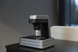
Grundium's compact Ocus whole slide scanners automate the process of digitizing microscope slides. The scanners produce high-resolution digital images of tissue and cell samples, which can then be accessed and shared online via a secure web browser. This eliminates the need for physical slide transport, reduces manual work, and enables remote collaboration among experts, streamlining the digital pathology workflow. Another distinct advantage of the Ocus scanners is the “live view” capability, which allows the pathologist to view and control a slide more rapidly than traditional scanning. These features enable WashU to provide the same expertise for frozen sections in the bi-state region as they do for telecytology.
The company's scanners are already in use across leading medical institutions in North America and Europe, with planned expansions into markets where physician shortages pose even greater challenges to healthcare access.
