Cedars-Sinai advances research that could aid early Alzheimer’s diagnosis
Three recently published studies from Cedars-Sinai investigators have deepened knowledge of how changes in the eye are linked to indicators of Alzheimer’s disease in the brain.
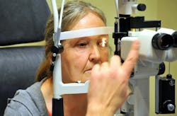
The eye-brain connection could help physicians diagnose patients with Alzheimer’s disease earlier, a key factor in developing effective treatments.
In a study published in the peer-reviewed journal Acta Neuropathologica Maya Koronyo-Hamaoui, PhD and fellow investigators compared retinal tissue from 45 patients diagnosed with Alzheimer’s-related cognitive impairment or dementia with tissue from 34 individuals with normal cognition or non-Alzheimer’s forms of dementia.
Investigators found that higher levels of abnormal tau in the retina corresponded to levels of tau in the brain, other brain changes related to Alzheimer’s disease, and cognitive decline.
In a study published in the peer-reviewed journal Acta Neuropathologica Communications, Koronyo-Hamaoui and co-investigators used leading-edge imaging and image-processing technology to compare amyloid plaques in the retinas of living patients who had early-stage cognitive impairment with those in individuals who had normal cognition.
Thirty-four patients underwent retinal and brain imaging, and cognitive testing. Analysis in 28 patients revealed two to three times as many plaques clustered near blood vessels in the retinas of patients with mild cognitive impairment or Alzheimer’s disease when compared with individuals with normal cognition. The numbers and position of the plaques correlated with cognitive decline and physical changes in the brain.
Koronyo-Hamaoui’s team also published a review article in the peer-reviewed journal Progress in Retinal and Eye Research that detailed additional Alzheimer’s disease biomarkers that have been identified in the retina.
These include reduced blood flow, deposits of amyloid-beta proteins inside blood vessel walls, damage to the barrier that prevents harmful substances from entering retinal tissue, inflammation, and damage to nerve cells.
“Imaging technology now being developed will allow us to see these changes in patients in clinical settings,” said Keith L. Black, MD, chair of the Department of Neurosurgery and the Ruth and Lawrence Harvey Chair in Neuroscience at Cedars-Sinai and co-author of these studies. “This technology, which is noninvasive and affordable, allows us to see changes in the cells and blood vessels in tremendous detail.”
Black and Koronyo-Hamaoui envision this technology as a tool to screen patients in primary care settings, with those who show features suggestive of Alzheimer’s disease referred for additional testing, such as a PET brain scan or cerebrospinal fluid or blood test. The technology could also assess disease progression and the effectiveness and safety of new treatments.

